Selected experience

Examples from my
pathology work

A selection of the studies and quantitative methods I have worked on across industry roles and independent consulting.

Publications and conference contributions

Oligonucleotide safety

ASO and siRNA programmes
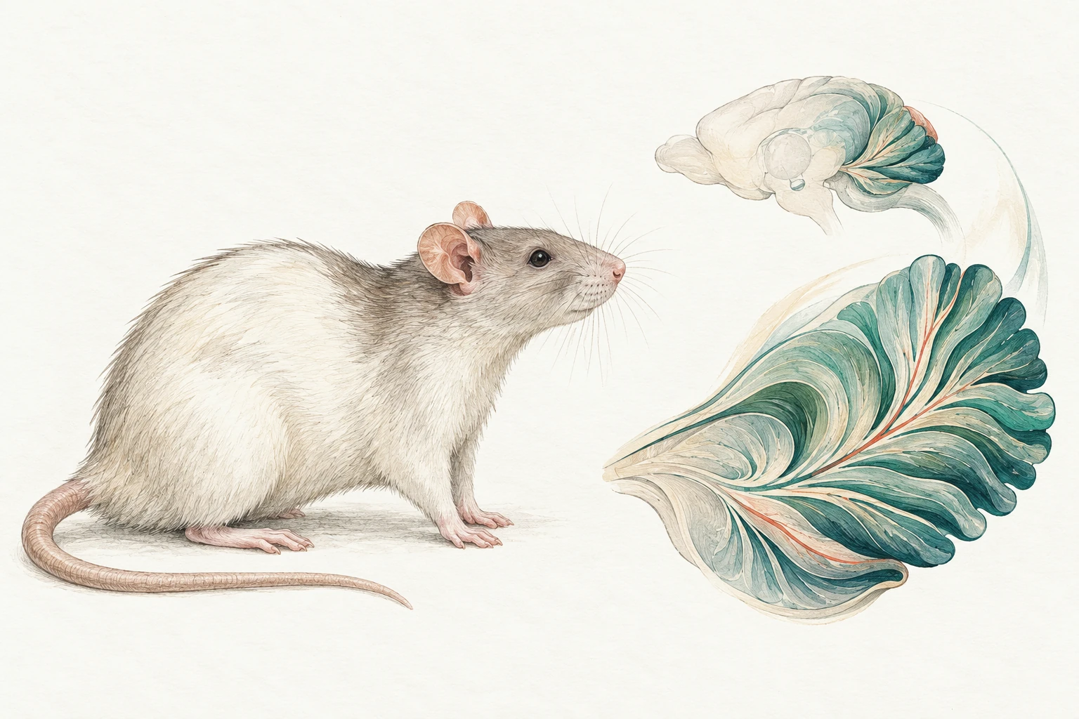
Editorial illustration of a laboratory rat beside a separate conceptual brain and cerebellar folia.

Central and peripheral nervous system assessment

My work includes primary reads and independent peer review of ASO and siRNA safety studies, including intrathecal programmes.

I interpret routine histology and ancillary stains alongside lesion distribution, tissue quality and other study observations.
H&E · IBA-1 · Calbindin · GFAP · Fluoro-Jade where available

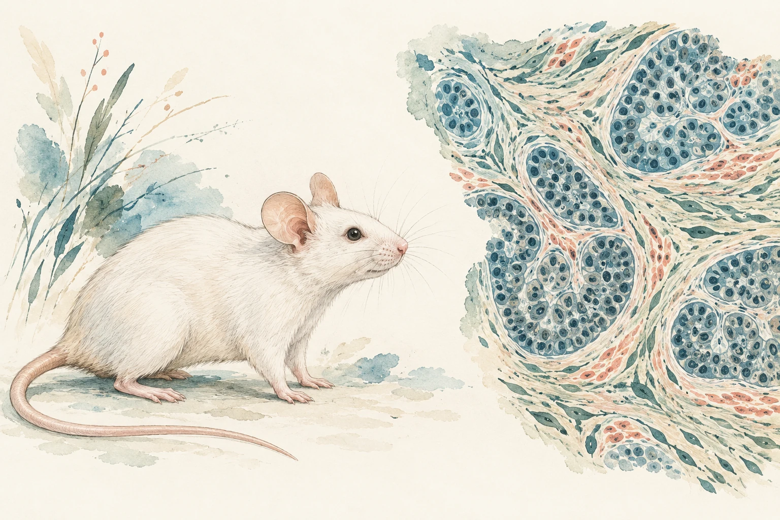
Oncology

Editorial illustration of a laboratory mouse beside painted epithelial nests and surrounding stroma.

Oncology and tissue-biomarker assessment

My oncology work has included tissue evaluation, IHC/ISH and image analysis, as well as collaboration with genomics teams on spatial methods.

Histology · IHC / ISH · Image analysis · Spatial-genomics collaboration

Oncology services

Disease and safety models

Explore model applications

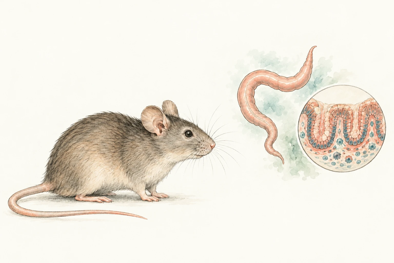
DSS-induced colitis · Mouse

Editorial illustration of a laboratory mouse beside a separate colon and epithelial crypt vignette.

DSS-induced colitis

Blinded histopathological scoring of colon Swiss rolls, including inflammation, epithelial injury, goblet-cell loss and crypt changes.

H&E · Defined histological scoring

mdx muscular dystrophy · Mouse

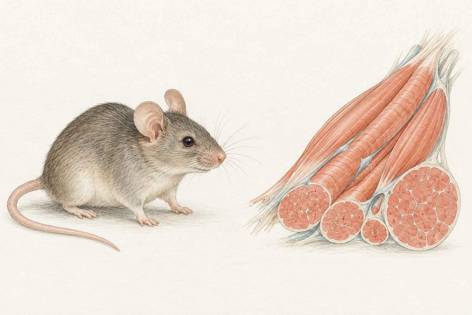
Editorial illustration of a laboratory mouse beside a conceptual skeletal-muscle fibre bundle.

Duchenne muscular dystrophy

Skeletal-muscle assessment combining evaluation of necrosis, regeneration and inflammation with myofiber-size measurements.

H&E · Lesion evaluation · Myofiber morphometry

Bleomycin-induced pulmonary fibrosis · Mouse

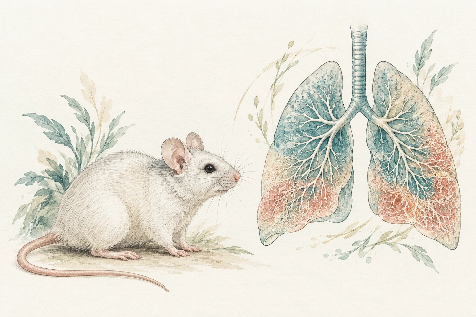
Editorial illustration of a laboratory mouse beside a conceptual lung and branching-airway vignette.

Pulmonary fibrosis

Assessment of collagen-stained lung sections using modified Ashcroft scoring across sampled fields.

Collagen staining · Semiquantitative fibrosis scoring

HDM-induced airway inflammation · Mouse

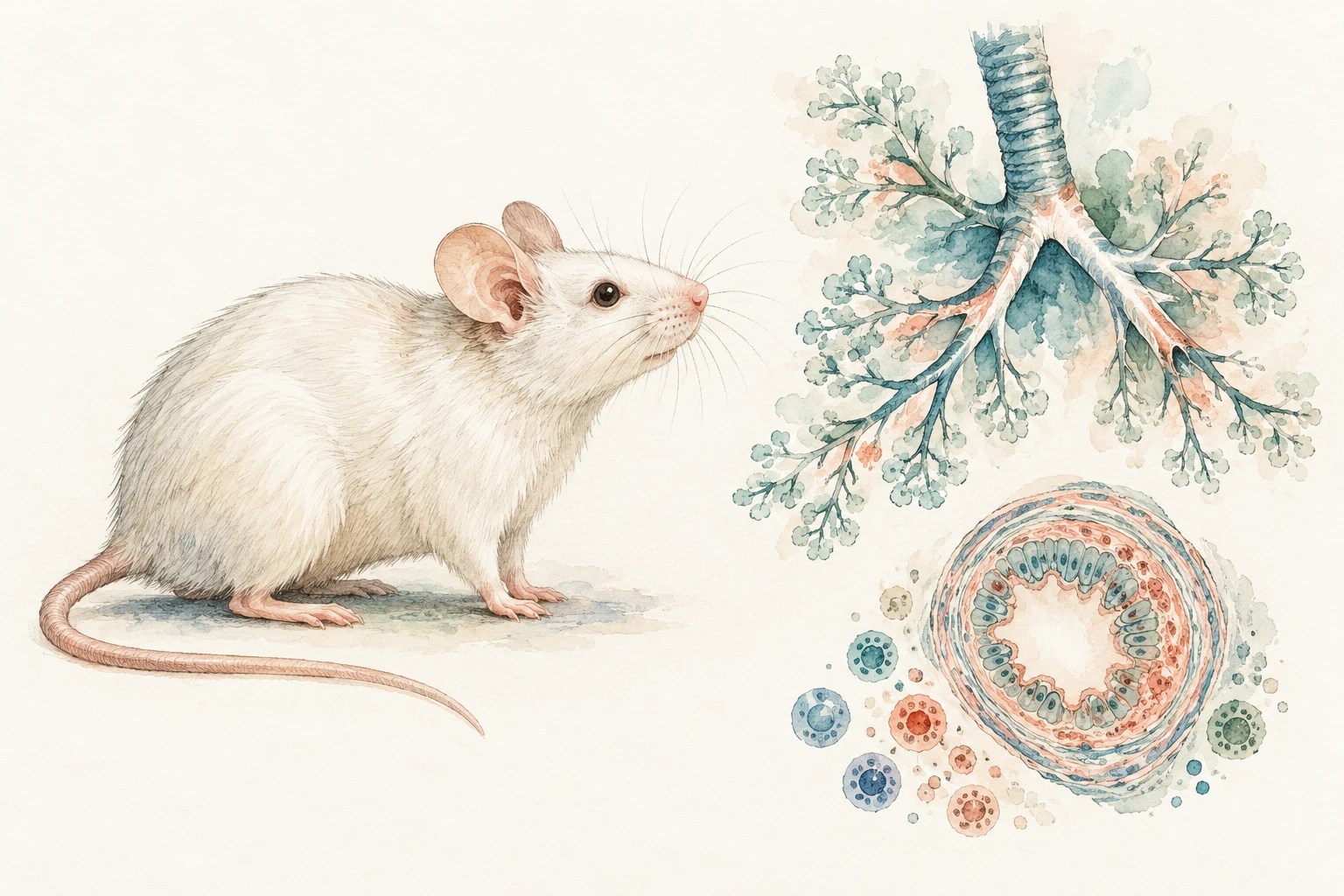
Ink and gouache illustration of a laboratory mouse beside separate branching airways and a simplified airway cross-section.

House-dust-mite airway inflammation

Evaluation of the distribution of airway inflammation and epithelial changes, with assessment of mast cells on ancillary-stained sections.

H&E · Toluidine blue · Inflammatory endpoints

Letrozole-induced PCOS · Mouse

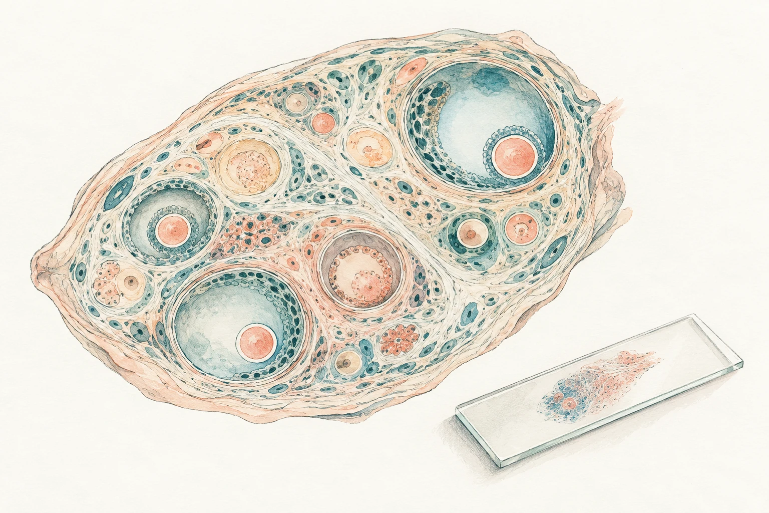
Ink and gouache illustration of an oval tissue vignette with follicle-like forms beside a glass slide.

Polycystic ovary syndrome

Pathological assessment in a letrozole-induced PCOS model, combining review of ovarian tissue sampling with adipose-tissue evaluation.

H&E · Tissue sampling · Adipose morphology

High-fat-diet model · Mouse

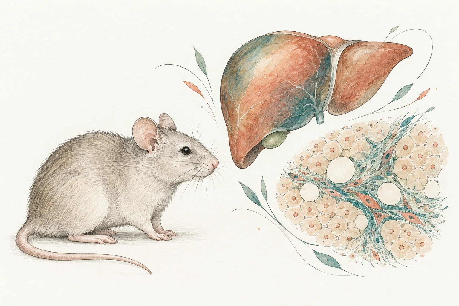
Editorial illustration of a laboratory mouse beside a separate liver silhouette and conceptual hepatic cells, lipid spaces and fibrous strands.

Fatty-liver model characterisation

Histopathological assessment of liver steatosis in a high-fat-diet mouse model, with review of the accompanying tissue changes.

Steatosis · Histopathology · Tissue review

β-amyloid model · Transgenic rat

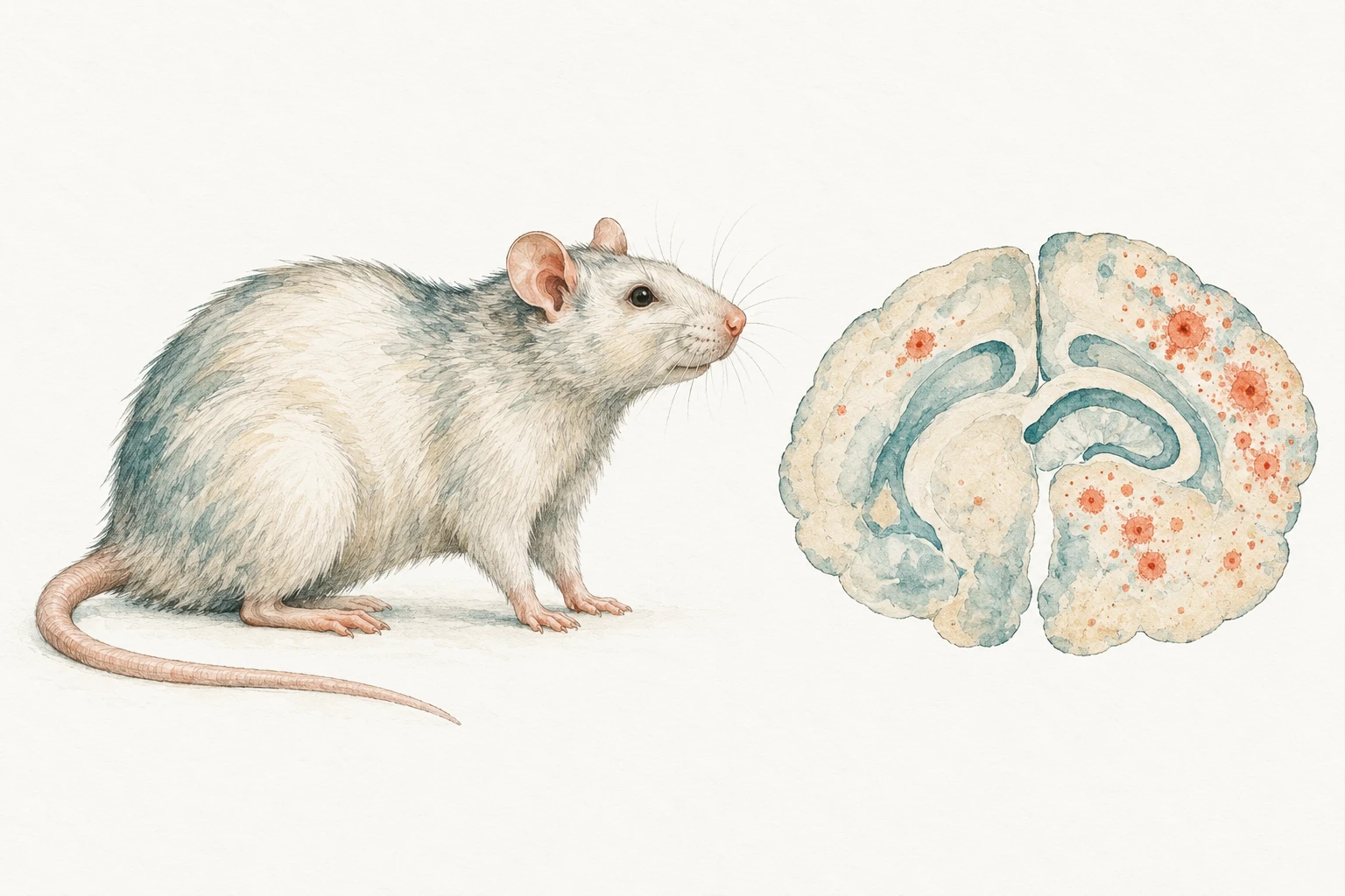
Ink and gouache illustration of a laboratory rat beside a separate brain-inspired vignette with scattered coral deposit-like marks.

β-Amyloid deposition

Regional assessment of β-amyloid immunolabelling in sagittal brain sections, with separate review of plaque distribution and intraneuronal signal.

IHC · Regional distribution · Semiquantitative assessment

Intracerebral AAV · Cynomolgus macaque

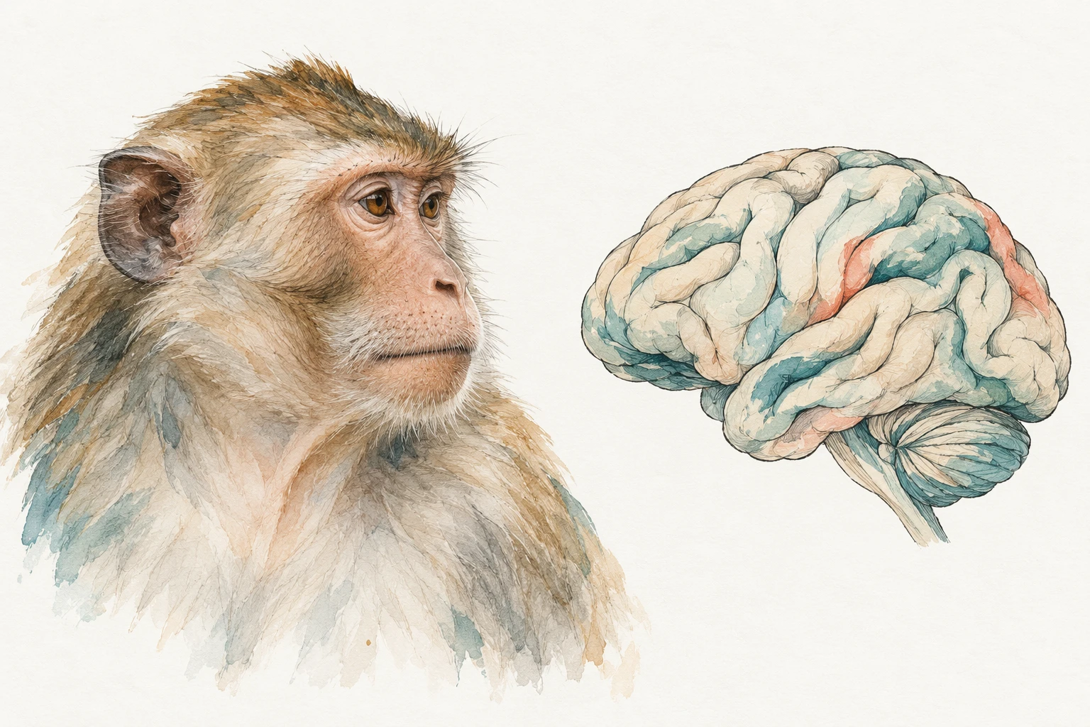
Ink and gouache illustration of a calm cynomolgus macaque beside a separate folded-brain-inspired vignette.

Vector delivery and regional neuropathology

Regional brain histopathology after intracerebral AAV administration, including assessment around delivery sites and surrounding tissue.

H&E · Regional neuropathology · Administration-site assessment

Ex vivo tissue models

Human skin explants · Ex vivo

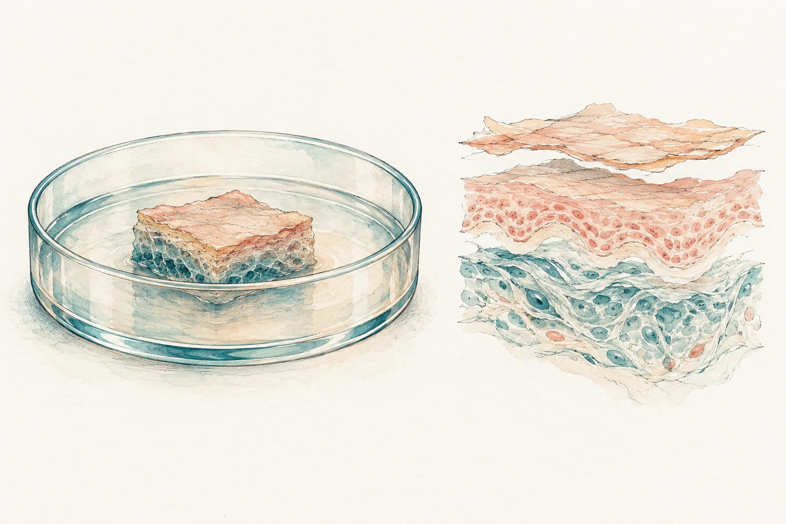
Ink and gouache illustration of a small skin-tissue explant in a shallow culture dish beside a layered tissue vignette.

Ex vivo human skin

Histological evaluation of skin architecture and integrity in human skin explants, supported by immunohistochemistry and epidermal-thickness measurements.

H&E · IHC · Epidermal morphometry

Image analysis

Quantitative methods combined with pathological review.

AI-assisted image analysis

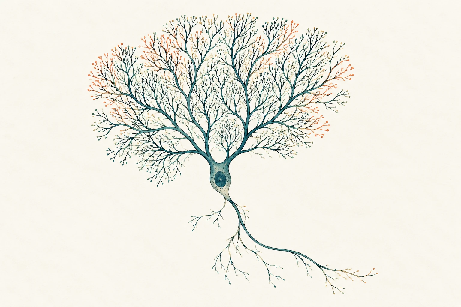
Ink and gouache illustration of a single fan-like branching Purkinje neuron on ivory paper.

Purkinje-cell quantification

Collaborative work on Purkinje-cell density in primate cerebellum. The workflow combined HALO AI tissue-layer segmentation and cell detection with QuPath processing and pathologist review.

H&E · HALO AI · QuPath · Expert review

Pathologist-directed histomorphometry

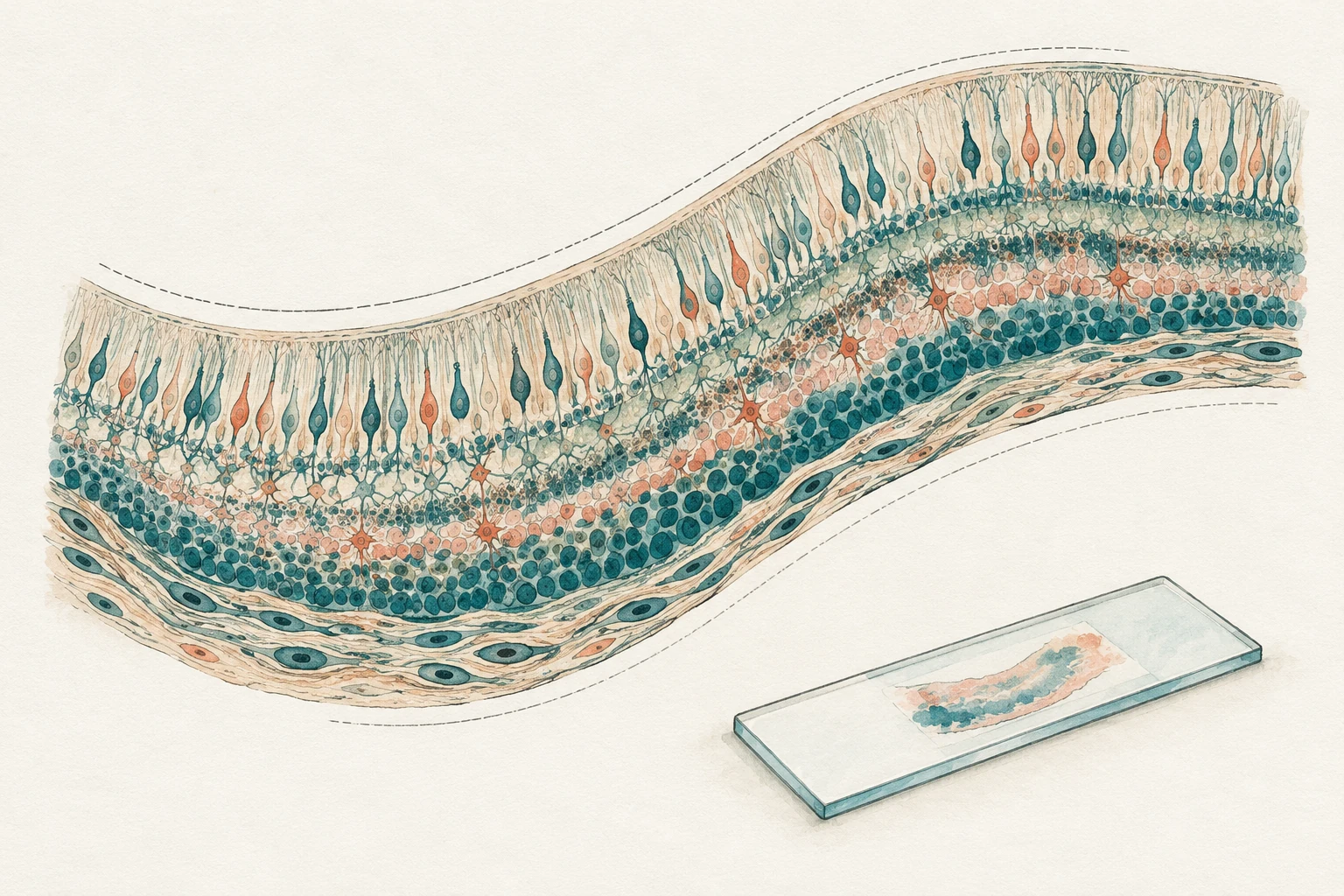
Hand-drawn layered retinal tissue concept with boundary traces and a glass microscope slide.

Retinal thickness measurements

Measurements on rat-eye whole-slide images at defined anatomical locations, with representative-section selection and review of tissue quality and section artefacts.

Whole-slide images · Anatomical sampling · Thickness measurements

Image-derived measurements

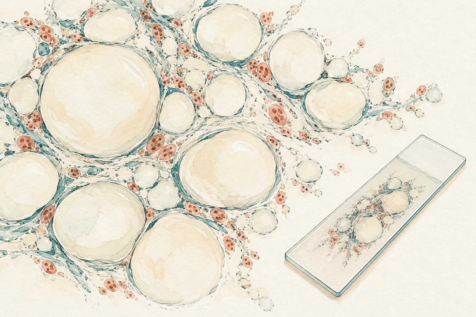
Ink and gouache illustration of rounded lipid-space-like profiles beside a microscope slide.

Adipose-tissue morphometry

Assessment of lipid-space area and diameter in a mouse disease model, combining image-derived measurements with review of sampling, segmentation and the underlying histology.

Lipid-space measurements · Sampling review · Histopathology

Contact

Discuss a study.

Email me the question, the material available and your key dates.